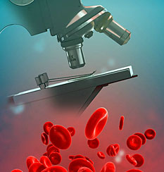
Advanced Microscopy
Feedsee Microscopy : Advanced Microscopy : Electron microscopes provide developers with sub-angstrom data for industrial applications
 Mexico's national research petroleum institute, the Instituto Mexicano del Petroleo (IMP), opened its center for advanced microscopy and research in 2006. In planning since 2002, the center features a complete suite of FEI tools that enable advanced nanoscale research and development. The opening was announced jointly by IMP and FEI Company. The state of the art center features FEI's XL 30 environmental scanning electron microscope (ESEM), a Nova 200 NanoLab, the first DualBeam installed in Latin America, two Tecnai transmission electron microscopes (TEMs), and FEI's flagship product, a Titan S/TEM, the world's most powerful commercially-available microscope. Researchers at IMP will use the FEI tools for a variety of applications including the study and development of catalysts for more efficient fuels, fuel cell membranes, and new nano-enabled materials such as anticorrosive products for oil refineries and pipelines, among others.
Evolution of Electron Microscopes
Electron microscopes, which use beams of electrons to form images, have made significant advancements since their invention in the 1930s. These improvements have increased resolution, improved image quality, expanded functionality, and enhanced our understanding of a wide variety of scientific phenomena. Here are some major improvements over the years:
- Increased Resolution: The resolution of electron microscopes has continually improved since their development. Modern high-end Transmission Electron Microscopes (TEMs) and Scanning Transmission Electron Microscopes (STEMs) can achieve atomic-scale resolution, enabling scientists to view individual atoms.
- Development of SEM: The invention of the Scanning Electron Microscope (SEM) was a significant advancement. Unlike TEMs, which produce a two-dimensional image, SEMs scan the surface of the specimen with a focused electron beam and produce three-dimensional images, providing topographical information.
- Better Electron Sources: The development of field emission electron sources has improved both the resolution and the quality of images. Field emission sources provide a more coherent, brighter source of electrons, allowing for better image clarity and resolution.
- Cryo-Electron Microscopy: Cryo-electron microscopy is a technique that involves rapidly freezing the sample to preserve its natural state and then observing it under an electron microscope. This technique has been revolutionary, especially in biology, where it is used to image proteins and other biological complexes in their natural state.
- Development of Analytical Techniques: Modern electron microscopes often come equipped with a variety of analytical tools. These include energy-dispersive X-ray spectroscopy (EDS) and electron energy loss spectroscopy (EELS), which can provide elemental and bonding information about the sample.
- Computer-Aided Image Acquisition and Processing: Computers have revolutionized data acquisition and processing in electron microscopy. They have allowed for the automation of many microscope functions, the digital recording and processing of images, and the reconstruction of 3D images from a series of 2D images.
- Environmental Electron Microscopy: The development of Environmental SEMs (ESEMs) allows for the imaging of samples in a more natural state (e.g., wet, gaseous), which was previously impossible due to the high vacuum requirements of traditional electron microscopes.
- Aberration Correction: Aberrations in the electron lenses can distort images and limit resolution. The development of aberration correctors, which correct these distortions, has greatly improved the resolution and image quality in both TEM and SEM.
- Time-Resolved Techniques: Ultrafast electron microscopes have been developed that can capture images with time resolutions on the picosecond and femtosecond scales, opening up the field of ultrafast electron microscopy.
Advancements in electron microscopy have dramatically enhanced its capabilities, enabling scientists to probe matter at previously unattainable resolutions and providing profound insights into the structure and behavior of materials and biological specimens.