Microscope Illumination
Feedsee Microscopy : Microscope Illumination : Consistent reflected lighting used a pre-centered mercury-fiber illuminator for epi-fluorescence
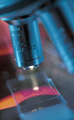 In 2006, Nikon's Intensilight mercury-fiber illuminator for bioscience and industrial microscopy applications was a pre-centered fiber illumination system with long lamp life and easy lamp replacement which never needs alignment. The Intensilight featured a no-flicker, stable light intensity which can be controlled with Nikon's NIS-Elements software. The illumination system provided quantitative fluorescence with outstanding transmission at lower UV wavelengths. Nikon's Intensilight included matched adapters for different families of Nikon microscopes. A built in open-close shutter made it easy to transfer the illumination source to multiple microscopes on the fly or to shutter the illuminator's light to the microscope specimen, and a neutral density wheel provides intensity control to prevent photobleaching of sensitive specimens while insuring shading free and extremely even specimen illumination.
Microscopy Lighting Techniques
There are several lighting techniques used in microscopy to illuminate the specimen and obtain clear images. These techniques often rely on different physical properties of light such as phase, polarization, and fluorescence. Here are some of them:
- Brightfield Illumination: This is the simplest form of lighting technique. It's called "brightfield" because it makes the field of view appear bright, with the specimens being dark. This is the most common form of lighting used in biological light microscopy.
- Darkfield Illumination: This technique uses a special condenser that directs light towards the specimen from the sides, rather than from directly beneath. This causes only the light scattered by the specimen to enter the objective lens, creating a "dark field" with a brightly lit specimen.
- Phase Contrast: This method makes use of the phase differences in light passed through different parts of a transparent specimen. Phase contrast illumination is especially useful for observing live cells and tissues, where staining may not be feasible.
- Differential Interference Contrast (DIC): Also known as Nomarski Interference Contrast, DIC uses polarized light and additional optical components to exaggerate the small differences in the index of refraction in different parts of the specimen. It results in a pseudo-3D, shadow-casted image, which is particularly useful for thick, unstained specimens.
- Polarized Light: Polarizing microscopy is a technique that uses polarized light to examine birefringent materials, such as crystals, polymers, and biological tissues like muscles. It can provide information about the composition and three-dimensional structure of these materials.
- Fluorescence: In fluorescence microscopy, the specimen is illuminated with a specific wavelength of light that excites fluorescent molecules (fluorophores), causing them to emit light of a longer wavelength. This emitted light is then collected to form an image. This technique is extremely sensitive and allows for specific labelling of different structures within a specimen.
- Confocal Microscopy: Confocal microscopy uses point illumination and a pinhole to exclude out-of-focus light, enhancing resolution and contrast. It is commonly used with fluorescence staining.
- Two-photon Excitation: This is a variant of fluorescence microscopy, where two photons of lower energy are absorbed simultaneously to excite the fluorophore. This technique provides better penetration depth and reduces photobleaching and phototoxicity compared to single-photon fluorescence microscopy.
Each of these techniques serves different purposes and provides unique advantages, and often they can be combined or used sequentially for a more comprehensive analysis of the specimen.